Peptide-Polymer Hybrids Repair Damaged Tissue at the Material/Tissue Interface
Hydroxyapatite-binding peptide (HABP) HABP has an inherent capacity to bind the calcium phosphate in dentin. Self-strengthening polymer with tethered HABP will diffuse into the mineralized tissue, in this case dentin, and the HABP will act as a scaffold for nucleation and growth of hydroxyapatite crystals. This activity will remineralize the damaged dentin and anchor the material (adhesive or varnish) to the dentin. Tethering the peptides to the polymer decreases the risk of bioactive-agent diffusion.
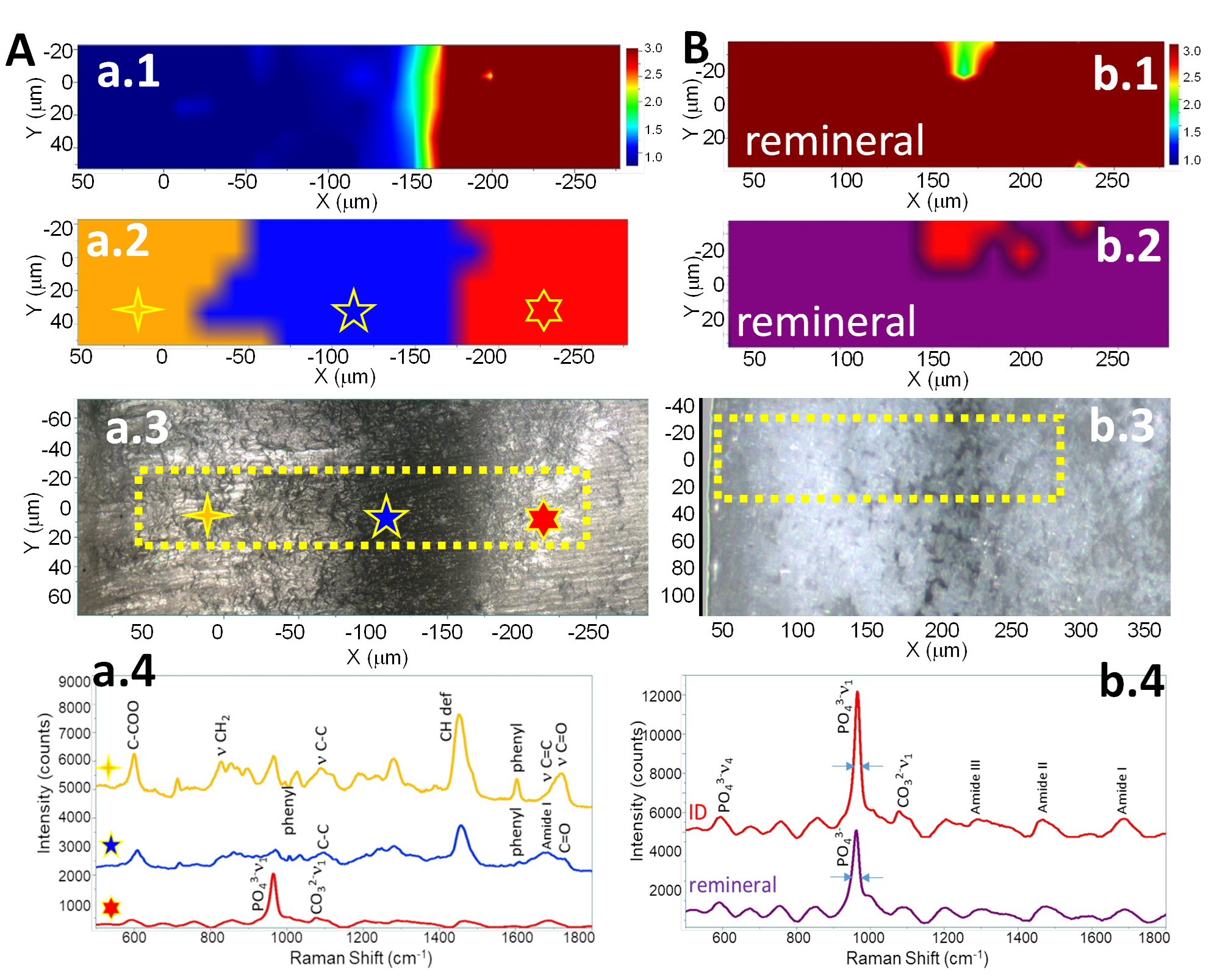
Fig. 7 RAMAN analysis of a/d interface before remineralization (A), and after remineralization (B). a.1 & b.1. Intensity ratio of mineral/collagen. a.2 & b.2 Intensity ratio of mineral/adhesive. a.3 & b.3 Visible images of the scanned areas for the chemical mapping. a.4. RAMAN spectra of HABP-tethered adhesive (Adh, Gold), adhesive-infiltrated demineralized dentin (AIDD, Blue) and intact dentin (ID, Red). b.4. RAMAN spectra of intact dentin (ID, top) and formed peptide-mediated remineral (bottom). FWHM of the P-O (960cm-1) is 19.8 & 23.7cm-1 for intact and remineralized dentin, respectively.
HABP-functionalized Adhesive at Interface with Dentin: Since micro-Raman spectroscopy (µRS) is non-destructive, the adhesive/dentin (a/d) interface was analyzed before (Fig. 7A) and after enzyme-mediated mineralization (Fig. 7B). Note: Blue denotes the lowest ratio and red the highest. Multi-variate data analysis was used to describe the chemical structure and spatial distribution of the components in the a/d interface. The intensity ratio of the P-O (mineral, 960)/Amide I (collagen, 1667) is presented in 7Aa.1 and 7Bb.1. The high intensity of the P-O band in 7Bb.1 indicates that the a/d interface is coated with mineral. A different region of the interface and the intensity ratio of the P-O (mineral, 960)/C-H (associated with adhesive, 1450) is presented in 7Aa.2 and 7Bb.2. The high intensity of this ratio indicates significant contribution from the P-O band―mineral coating the a/d interface (purple, 7Bb.2). The visible image (7Aa.3) represents the scanned area presented in 7Aa.2 and the corresponding spectra are presented in 7Aa.4. The visible image (7Bb.3) represents the scanned area presented in 7Bb.2. A comparison of the full-width half-maximum (FWHM) of the P-O band in intact dentin and peptide-mediated remineralized dentin reveals minor differences in the mineral (7Bb.4).
Publications:
Ye Q, Spencer P, Yuca E, Tamerler C. Engineered Peptide Repairs Defective Adhesive-Dentin Interface. Macromolecular Materials and Engineering. 302:1600487, 2017, DOI: 10.1002/mame.201600487
Spencer P, Ye Q, Song L, Parthasarathy R, Boone K, Misra A, Tamerler C. Threats to Adhesive/Dentin Interfacial Integrity and Next Generation Bio-enabled Multifunctional Adhesives. J Biomed Mater Res Part B:107B:2673-2683, 2019
Paulette Spencer, Qiang Ye, Nilan J. B. Kamathewatta, Sarah K. Woolfolk, Brenda S. Bohaty, Anil Misra, and Candan Tamerler. Chemometrics-Assisted Raman Spectroscopy Characterization of Tunable Polymer-Peptide Hybrids for Dental Tissue Repair. Frontiers in Materials 8:681415, 2021.
Esra Yuca, Sheng-Xue Xie, Linyong Song, Kyle Boone, Sarah K. Woolfolk, Nilan Kamathewatta, Philip Elrod, Qiang Ye, Paulette Spencer, and Candan Tamerler. Reconfigurable Dual Peptide Tethered Polymer System Offers a Synergistic Solution for Next Generation Dental Adhesives. International J of Molecular Sciences 22:6552, 2021