Multi-Modal and Multi-Scalar Analyses of Hierarchically Organized Tissues
Tissues and organs are by nature complex, hierarchically organized composites that exhibit multi-scale structure/property interdependence. The interfaces in biological systems that exist at the transition from one tissue-type to another are characterized by vastly different structures and functions and are hierarchically structured to provide optimal functionality. IBER researchers have developed capabilities and expertise for investigating multi-scalar characteristics through the application of multi-modal techniques.
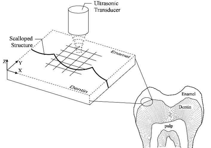
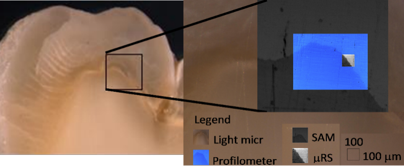
The dentino-enamel junction (DEJ), which represents a biological interface that successfully joins stiff, brittle enamel to compliant, relatively ductile dentin, has been widely cited as an example of a material system in which structural hierarchy contributes significantly to the longevity and performance of the tooth. Cracks in the enamel fail to propagate through the DEJ to the subjacent dentin, but the underlying reasons for this property remain unclear. The identification of strategic elements that mitigate damage and crack propagation between enamel and dentin requires systematic multi-modal and multi-scalar analyses, and using those results to predict behavior requires mathematical models. IBER laboratories are equipped with the instrumentation and expertise necessary to tackle such questions, including the capability of taking diverse homotopic measurements (precisely aligned across different methods and scales, Figures 1 and 2) and using those measurements to develop mathematical models that explore how features observed at different scales give rise to the sample’s unique properties. Indeed, the research team working with Dr. Misra has used homotopic measurements to develop models that explore stress transfer within the DEJ.
Publications:
Marangos O, Misra A, Spencer P, Bohaty B, Katz JL. Physico-mechanical properties determination using microscale homotopic measurements: application to sound and caries-affected primary tooth dentin. Acta Biomater 2009;5(4):1338-48
Marangos O, Misra A, Spencer P, Katz JL. Scanning acoustic microscopy investigation of frequency-dependent reflectance of acid- etched human dentin using homotopic measurements. IEEE Trans Ultrason Ferroelectr Freq Control 2011;58(3):585-95
Misra A, Marangos O, Parthasarathy R, Spencer P. Micro-scale analysis of compositional and mechanical properties of primary tooth dentin using homotopic measurements. In: Lacoviello D, Andreaus U, editors. Micro-scale analysis of compositional and mechanical properties of primary tooth dentin using homotopic measurements. Dordrecht, The Netherlands: Springer; 2013.
Misra A, Marangos O, Parthasarathy R, Spencer P. Micro-scale analysis of compositional and mechanical properties of dentin using homotopic measurements. Biomedical Imagine and Computational Modeling in Biomechanics 2013;4:131-141
Misra A, Marangos O, Spencer P. Dentinoenamel junction: motif for interfacial mechanics of dissimilar materials. In: Spencer P, Misra A, editors. Material-Tissue Interfacial Phenomena. Amsterdam: Woodhead Publishing; 2017. p 267-281.