Imaging Equipment
The instrumentation within the Institute for Bioengineering Research is used for both research and teaching. The following instruments are included at the Institute for Bioengineering Research and are located in Learned Hall. To inquire about training and instrument use at the Institute for Bioengineering Research Laboratories, please complete the IBER Request Form. A completed IBER Request Form must be on file for anyone working in the laboratories. For more information contact:
Qiang (Charles) Ye, Ph.D.
Director, Institute for Bioengineering Research Laboratories
Office: Learned Hall 5101E
Phone: 785-864-1746
E-mail: yeq@ku.edu
C-SAM Gen 5 System Scanning Acoustic Microscopes (SAM)
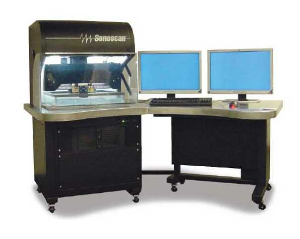 C-SAM Gen 5 system Scanning Acoustic Microscopes (Sonoscan, IL) comes with a set of 4 high precision acoustic lenses: 15, 30, 100 and 300 MHz, associated optics, computer and proprietary software. The C-SAM Gen 5 system consists of dual high frequency, high power, digital pulser/receiver for use with ultrasonic transducers up to 400 MHz, a x-y-z positioning system with 0.5 µm precision, a temperature controlled bath, capability to perform thru-scan measurements and capability to provide raw data for custom processing. Additional custom designed transducers will be acquired to the proposed R01. Acoustics lab is also equipped with the following: Textronix TDS5104 Digital Oscilloscope (1 GHz, 4 Channel), GE-Panametrics 5900 Pulser Receiver (200 MHz) and Paired Set of Ultrasonic Transducers each (cable connectors): Longitudinal @: 100 KHz; 500 KHz; 2.25 MHz; 5 MHz; 10 MHz; 20MHz, Shear @: 100 KHz; 500 KHz; 2.25 MHz; 5 MHz; 10 MHz; 20MHz.
Xradia MicroXCT Tomographic X-Ray Microscope
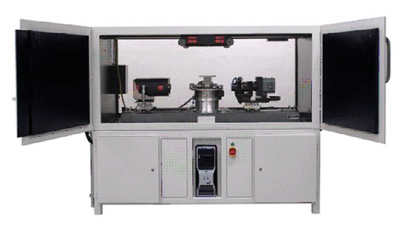 The specifications of this tomographic X-ray microscope include a 90 keV microfocused source, 2K x 2K CCD camera, multiple resolution modes down to 400 nm voxels, Phase Enhanced detectors and soft tissue imaging of samples up to 100 mm in diameter, and a precision stage resting on granite base. It uses phase contrast, instead of absorption contrast, and offers substantially improved image contrast for soft biological tissues to allow their visualization. The novel detector of the MicroXCT-400 with a pixel resolution of about 0.3 mm and spatial resolution of less than 1 mm has the highest performance of any commercially available x-ray detector and provides powerful 3D visualization capability to biomedical researchers. The Xradia Micro XCT has a large chamber and a heavy loading precision stage can bear the sample and/or in situ fixtures weighing up to 15 kg mounted onto a granite block. It also allows in situ thermal mechanical tests to be conducted over long periods of time, without being affected by mechanical vibration and temperature fluctuations. Additionally, Xradia’s systems come complete with workstation for fast tomography reconstruction, image processing, segmentation and analysis.
Optical Digital Image Capture System
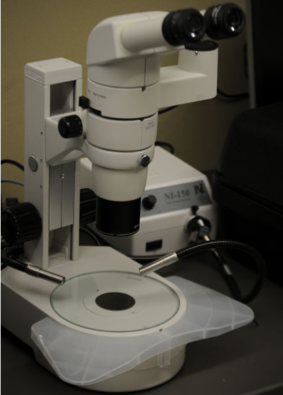
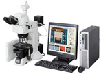 Nikon LV100DU brightfield, darkfield episcopic/diascopic microscope equipped with CFI LU Plan Fluor EPI objectives and Plan Apo 100x Oil objectives with high NA / working distance and a Nikon SMZ800 stereo microscope with 1.0x-6.3x variable zoom; either could be coupled to a 5MP cooled color CCD camera and an advanced imaging station.